Immunoassays – such as ELISA and ELISpot – are indispensable tools for immunology research. They work based on principles of antibody-epitope interaction, providing critical insights into the inter- and intracellular signalling pathways that drive biological processes. They hold applications in investigating: the immune response to pathogens, the progression of autoimmune disease, the efficacy of vaccines, and organ post-transplant success.
Of this assay type, the Enzyme-Linked Immunosorbent Assay or “ELISA” is perhaps the most well-known. It is widely used in routine laboratory settings, including biosafety level 2 (BSL-2) environments, and is a go-to method for detecting and quantifying protein-based analytes such as cytokines, antibodies, and antigens. Yet, despite its widespread application and versatile utility, the ELISA assay is not a ‘one-size-fits-all’ solution, especially in immunology research.
Immunology is a complex, nuanced field in which rare events and intricate cell maturation patterns necessitate highly sensitive and specific research methods. In the right application setting, ELISpot can offer greater resolution and more scientifically valid results. This article highlights some of the key similarities and differences between ELISpot vs ELISA assays. It also aims to equip researchers with the knowledge required to confidently select the most appropriate assay for their immunology application.
ELISA is a plate-based assay system designed for the detection and quantification of specific antigens or antibodies within a liquid sample. They rely upon the enzyme-linked detection methods – typically mediated by reporter enzymes – to generate a quantifiable signal (typically colorimetric, but sometimes chemiluminescent or fluorescent) proportional to the abundance of a target analyte. There are several types of ELISA assay kits, each offering benefits depending on their application. These include direct, indirect, sandwich and competitive methods.
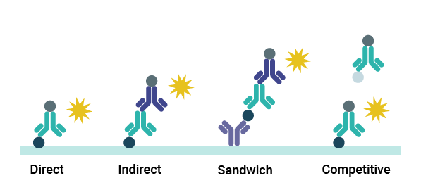
In most ELISA methods, a specific antigen or capture antibody is first immobilized on the plastic surface of a microplate well. Liquid sample containing the target analyte is then added before the plate is incubated (temperature and duration variable), allowing the target time to bind to the immobilized component. When the incubation time is up, any unbound target is washed away using a non-reactive buffer, such as PBS, TBS or Tween-20. A detection antibody is then introduced.
Primary detection antibodies are either directly conjugated to a reporter enzyme or bound to the reporter by an enzyme-linked secondary. The reporter produces a quantifiable signal that can be analyzed by a microplate reader when a specific substrate (such as alkaline phosphatase) is added. The intensity of this signal is directly proportional to the concentration of the target analyte in the liquid sample.
ELISpot assays are another example of an enzyme-linked immunoassay. Unlike traditional ELISAs, ELISpot assays are precisely designed to detect and quantify the number of individual cells secreting a target protein. While they share many similarities with the ELISA method, it’s important to remember that ELISpot protocols and applications are distinct.
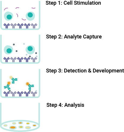
Performed in 96-well or 8-well strip microplates with specialized membranes, ELISpot assay kits utilize a sensitive sandwich-based detection approach to achieve target detection. Many suppliers offer both pre-coated and non-coated microplate options to allow researchers flexibility in their experimental design.
During an ELISpot assay, a known number of viable cells are added to each microplate well containing a membrane pre-coated with capture antibodies. The plate is then incubated (typically for 1-2 days), during which the cells secrete the target analyte. These secreted molecules are immediately captured by the antibodies immobilized directly beneath each cell, resulting in localized analyte deposition. After the incubation period, the cells are carefully washed away, leaving only the secreted bound analyte. Detection antibodies are then added to form a sandwich complex with the captured protein, followed by an enzyme-linked reporter and a chromogenic substrate (such as horseradish peroxidase or alkaline phosphatase). The enzymatic reaction generates visible coloured spots, each representing a single analyte-secreting cell. These can be quantified using microscopes or specialized ELISpot readers.
At first glance, ELISpot and ELISA assay kits appear remarkably similar – both harness sophisticated antibody-based reporting and rely on the immobilization of reagents within microplate wells. However, while they share the same core molecular principles, ELISpot and ELISA assay kits are engineered to interrogate fundamentally different aspects of immune biology.
While ELISA assays are designed to quantify the total concentration of an analyte present in a liquid sample—like serum, plasma, or cell culture supernatant—ELISpot offers a distinct advantage when it comes to profiling immune function. It allows researchers to analyze and quantify the distinct number of individual cells actively secreting a target molecule, providing detailed insights into T cell behaviour and cytokine expression.
To further highlight the differences between ELISpot and ELISA assays, the key characteristics of both are summarized in the table below.
| Feature | ELISA | ELISpot |
| Analyte Detection | Total concentration within a liquid sample | Number of individual, analyte-secreting cells |
| Sensitivity | Less sensitive | More sensitive |
| Resolution | Not single-cell level | Single-cell level |
Choosing an ELISpot assay is a great first move towards innovation in your immunology workflow. Their single-cell resolution, sensitivity to rare cell sensitivity and constant ongoing technological evolution allow them to meet the high standards of modern immunology research, while building on the familiar principles of ELISA-based workflows. ELISpot assay kits are the perfect choice for researchers looking to identify disease states at early stages, detect low-abundance pathogen stimulation or explore low-level inflammatory states. They can be used together, alongside ELISA and flow cytometry methods, to offer an additional perspective on immune cell function.1
In summary, choose an ELISpot assay when:
ELISpot has been used extensively in vaccine efficacy testing for many years.23 In fact, they are instrumental in quantifying T cell responses post-vaccination, informing novel prophylactic development and re-dosing strategies. By providing insights into T cell-mediated immune responses, ELISpot enable scientists and clinicians to accurately evaluate vaccine success, monitor long-term immunity, and make informed decisions on public health strategies and therapeutic interventions. Their inherent sensitivity also makes ELISpot assays ideal for infectious disease research, enabling the detection of low-frequency, pathogen-specific immune responses. A prime example is their crucial role in tuberculosis (TB) research: their ability to detect and differentiate between the T cell responses of patients with latent versus active TB infection has led to significant applications not only in laboratory research but also in the clinic.456
In the field of cancer immunotherapy research, ELISpot also helps monitor patient responses. It allows researchers to quantify patient responses to novel treatments, such as CAR-T cell therapy, by quantifying tumor-specific T cell activation. Furthermore, it plays a vital role in autoimmune disease research, supporting researchers in the profiling cytokine expression and T cell dysregulation. Some experts believe that ELISpot may also one day support clinicians in the diagnosis of hard-to-stratify post-acute inflammatory syndromes such as Long COVID or myalgic encephalomyelitis (ME/CFS); while this application remains in development, it promises to revolutionize point-of-care diagnostics if regulatory approval is achieved.
Virax Biolabs offers a variety of ELISpot kits and services for research use. We offer both coated and non-coated microplate options designed to target a variety of epitopes with the highest degree of specificity. In addition, we can work with you to support custom assay design, utilizing the latest peptide pool technology to achieve accurate T cell stimulation reflective of your research needs. Our ELISpot offerings are guided by expert immunology knowledge, industry expertise and experience in novel in vitro diagnostic (IVD) assay development (ongoing).
**
**